Long Bone Diagram Endosteum - 1 Schematic Drawing Of A Longitudinal Section Through A Long Bone Download Scientific Diagram / Bone marrow is found in the bone cavities of long bones and is involved in the production of blood cells.
Long Bone Diagram Endosteum - 1 Schematic Drawing Of A Longitudinal Section Through A Long Bone Download Scientific Diagram / Bone marrow is found in the bone cavities of long bones and is involved in the production of blood cells.. • internal bone surfaces are covered with a delicate connective tissue membrane known as the endosteum. Of long bones, and epiphyseal ends of bones. They are one of five types of bones: The endosteum (plural endostea) is a thin vascular membrane of connective tissue that lines the inner surface of the bony tissue that forms the medullary cavity of long bones. Figure 6.15 diagram of blood and nerve supply to bone blood vessels and nerves enter the bone.
The elongated, cylindrical shaft of long bone that ossifies from the primary centre of ossification. Bone tissue mainly consists of bone cells (osteoblasts, osteocytes, and osteoclasts) and a mineralized extracellular matrix that is primarily made up of regulate bone remodeling. This endosteal surface is usually resorbed during long periods of malnutrition, resulting in less cortical thickness. Long, short, flat, irregular and sesamoid. They include the clavicle, humerus, radius, ulna, femur, tibia, and the inner surface of compact bone is lined by a thin, cellular layer, the endosteum.
Answered Long Bone Fill In The Labels On The Bartleby from prod-qna-question-images.s3.amazonaws.com This layer of membrane envelopes the spongy tissue, the medullary cavity and the endosteum mainly aids in bone growth, repair and remodeling whereas, periosteum aids bone sensitivity and nourishment along with the above activities. It is made up of compact bone and • marrow cavity: In an adult, most red blood cells are formed in the marrow in flat bones. The blue represents additional matrix filling in the space btwn. Want to learn more about it? The elongated, cylindrical shaft of long bone that ossifies from the primary centre of ossification. Long bones are those in which the length exceeds the breadth and thickness. Blood vessels and tissue surrounding the injured area bleed and if medullary lesions develop along the inner aspect of the cortical bones, especially in the long bones, endosteal scalloping may be observed.
.each long tubular bone of the limbs presents a cylindrical cavity named marrow cavity and it is lined with the medullary membrane called endosteum.
Cortical bone appears radiopaque (white) on radiographs as the outermost layer of bone. The diaphysis and the epiphysis (figure 6.3.1). • the long and short hones are formed externally of compact bone, but their endosteums are irregular due to presence of spongy bone. Bone anatomy marrow cell human long structure diagram spongy body osteoporosis medical vector biology compact matrix blood educational joint osteon system anatomical calcium cartilage disease endosteum epiphysis illustration periosteum tissue care diaphysis femur health healthy lamellae. Osteoclasts on the inside in the endosteum remove this bone to maintain the bone diameter. Deep to the cortex is the medullary cavity. The circumferential lamellar bone resists compressive forces. Both the periosteum and the. □ red bone marrow □ white bone marrow. The endosteum is located on the internal surface of the bone, being the membranous layer that covers the medullary cavity, the bony trabeculae (spongy part of the bone), the haversian canals and internal walls of the compact long bones. The elongated, cylindrical shaft of long bone that ossifies from the primary centre of ossification. The long bones are those that are longer than they are wide. Long, short, flat, irregular and sesamoid.
Bone marrow is found in the bone cavities of long bones and is involved in the production of blood cells. Learn with flashcards, games and more — for free. Mesenchymal progenitors were isolated and identified. Of long bones, and epiphyseal ends of bones. Osteoclasts on the inside in the endosteum remove this bone to maintain the bone diameter.
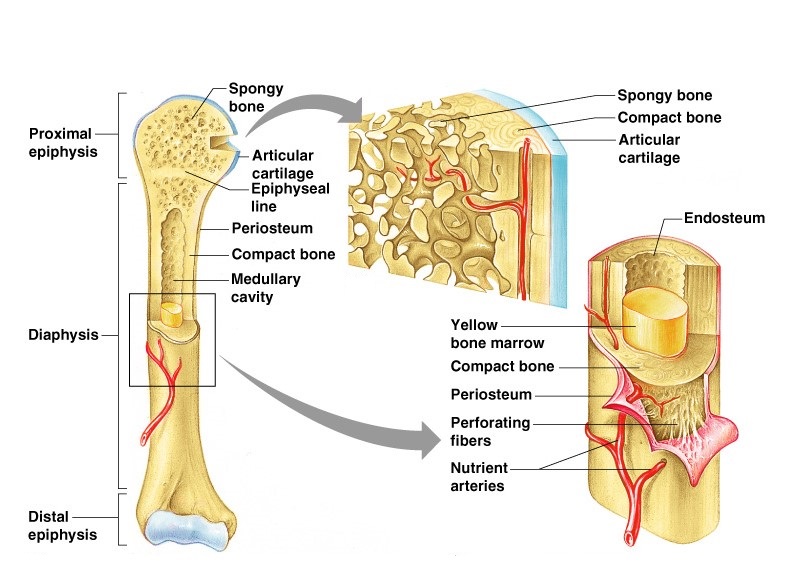
Bone Anatomy from cdn.thinglink.me The diaphysis is the hollow, tubular shaft that runs between the proximal and the osteoblast is the bone cell responsible for forming new bone and is found in the growing portions of bone, including the endosteum and the. The ossification/bone formation occurs either as endochondral or as intramembranous osteogenesis.the difference lies in the presence of bone formation: The long bones are those that are longer than they are wide. Periosteum and endosteum the external surface of bone is covered by the periosteum and its internal surface is lined by the endosteum. Long bones, especially the femur and tibia, are subjected to most of the load during daily activities and they are crucial for skeletal mobility. Parts of long bone (applies to other bones too). The diaphysis and the epiphysis (figure 6.3.1). The delicate connective tissue layer lining the inside surface of compact bone.
The end of the long bone is the epiphysis and the shaft is the diaphysis.
Blood vessels and tissue surrounding the injured area bleed and if medullary lesions develop along the inner aspect of the cortical bones, especially in the long bones, endosteal scalloping may be observed. • the long and short hones are formed externally of compact bone, but their endosteums are irregular due to presence of spongy bone. The blue represents additional matrix filling in the space btwn. Long bones are those that are longer than they are wide. Long bones, especially the femur and tibia, are subjected to most of the load during daily activities and they are crucial for skeletal mobility. A long bone has diaphyseal bone is organized to create the best balance between weight and structural strength. • internal bone surfaces are covered with a delicate connective tissue membrane known as the endosteum. (a) the schematic diagram of isolating mps from different regions of rat long bones. In an adult, most red blood cells are formed in the marrow in flat bones. The diaphysis and the epiphysis. Both the periosteum and the. Long bones are those in which the length exceeds the breadth and thickness. The endosteum can be seen in the t.s.
Endosteum and periosteum contribute to bone repair and reconstruction after a fracture occurs. It is found in bones such as the humerus and the. Make sure that you follow all the guidelines for biological drawings: This endosteal surface is usually resorbed during long periods of malnutrition, resulting in less cortical thickness. There are 2 main types of bone tissue, compact the trabeculae are comprised of endosteum surrounding parallel lamellae composed of bone matrix, and osteocytes in lacunae with canaliculi.
Structure And Functions Of Bones Online Science Notes from onlinesciencenotes.com Cancellous bone is remodeled by endosteum. Long bones, especially the femur and tibia, are subjected to most of the load during daily activities and they are crucial for skeletal mobility. Parts of long bone (applies to other bones too). Structure of long bone although there are many different types of bones in the skeleton, we will endosteum: At the ends of the bone the periosteum is continuous with the joint. Long bones are formed from a cartilage model precursor by endochondral ossification (see the image below) and can range in size from a phalanx to a femur. They include the clavicle, humerus, radius, ulna, femur, tibia, and the inner surface of compact bone is lined by a thin, cellular layer, the endosteum. It is found in bones such as the humerus and the.
Cells were isolated from the above figure 1.
It is made up of compact bone and • marrow cavity: The ossification/bone formation occurs either as endochondral or as intramembranous osteogenesis.the difference lies in the presence of bone formation: Of long bones, and epiphyseal ends of bones. The bones in your body have 3 major types of bone cells. Long bones, especially the femur and tibia, are subjected to most of the load during daily activities and they are crucial for skeletal mobility. The diaphysis and the epiphysis (figure 6.3.1). When osteoclasts start removing less bone, or osteoblasts start adding more bone, the. 14 (makes up the whole area). The endosteum appears at the interface between the. • the long and short hones are formed externally of compact bone, but their endosteums are irregular due to presence of spongy bone. It is best visualized in long bones. Bone tissue mainly consists of bone cells (osteoblasts, osteocytes, and osteoclasts) and a mineralized extracellular matrix that is primarily made up of regulate bone remodeling. Osteoclasts on the inside in the endosteum remove this bone to maintain the bone diameter.
Long bones are those that are longer than they are wide long bone diagram. This endosteal surface is usually resorbed during long periods of malnutrition, resulting in less cortical thickness.


0 Comments